介绍
缺铁性贫血。
患者数据
年龄:60岁
性别:女
CT
壶腹部周围胃肠道间质瘤 (GIST)
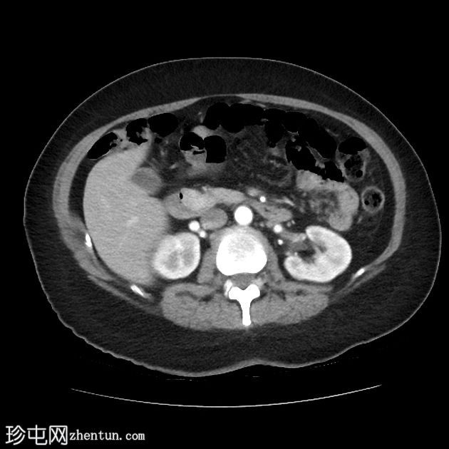
Axial C+
arterial phase
壶腹部周围胃肠道间质瘤 (GIST)
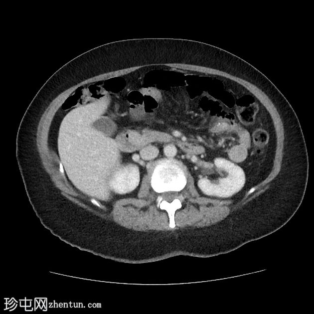
Axial C+ portal
venous phase
壶腹部周围胃肠道间质瘤 (GIST)
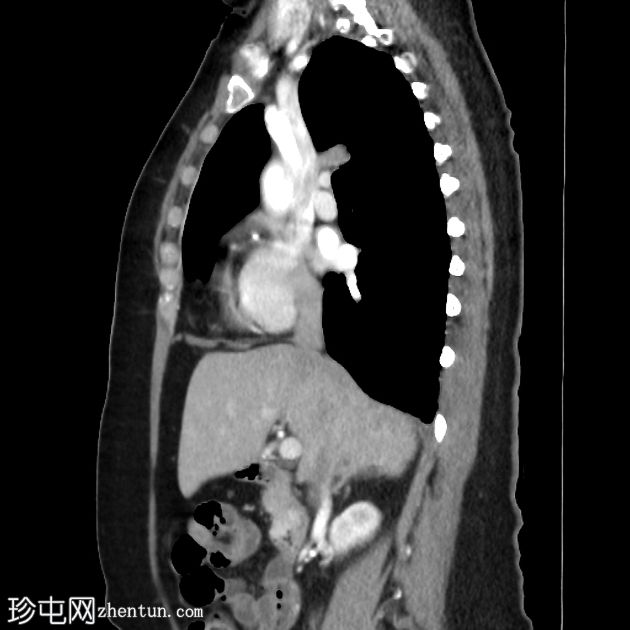
Sagittal C+
arterial phase
壶腹部周围胃肠道间质瘤 (GIST)
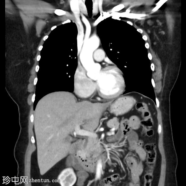
Coronal C+
arterial phase
动脉增强肿块位于胰头/十二指肠第二部分,位于壶腹部前方。无胰腺或胆管扩张。无淋巴结肿大。
MRI
壶腹部周围胃肠道间质瘤 (GIST)
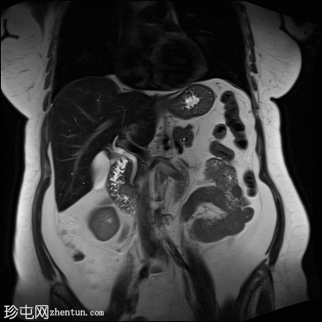
Coronal
T2
壶腹部周围胃肠道间质瘤 (GIST)
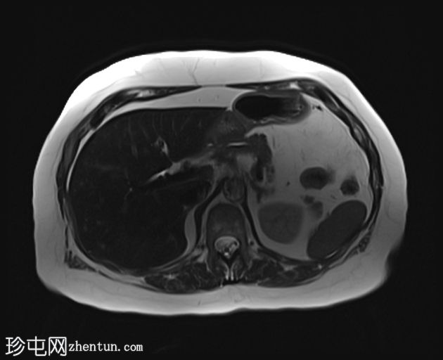
Axial
T2
壶腹部周围胃肠道间质瘤 (GIST)
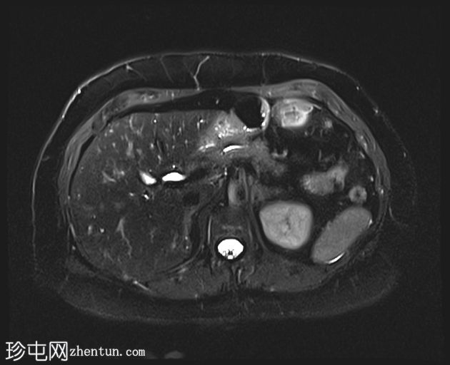
Axial T2
fat sat
壶腹部周围胃肠道间质瘤 (GIST)
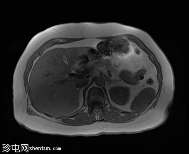
Axial T1
in-phase
壶腹部周围胃肠道间质瘤 (GIST)
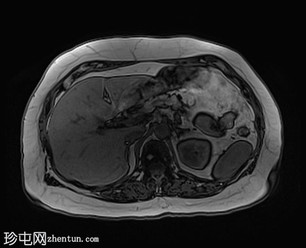
Axial T1
out-of-phase
壶腹部周围胃肠道间质瘤 (GIST)
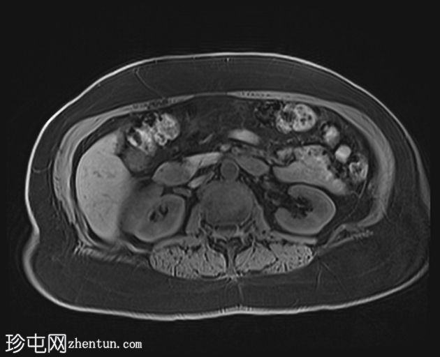
Axial T1
fat sat
壶腹部周围胃肠道间质瘤 (GIST)
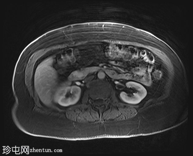
Axial T1 C+
fat sat (35s)
壶腹部周围胃肠道间质瘤 (GIST)
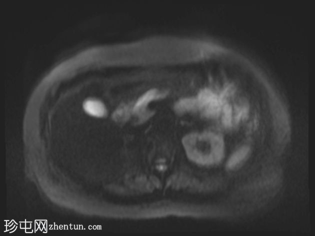
Axial DWI
(B800)
壶腹部周围胃肠道间质瘤 (GIST)
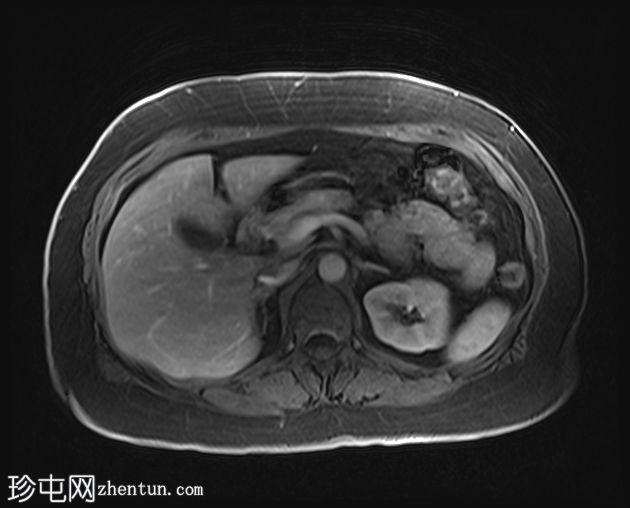
Axial T1 C+
fat sat (70s)
壶腹部周围胃肠道间质瘤 (GIST)
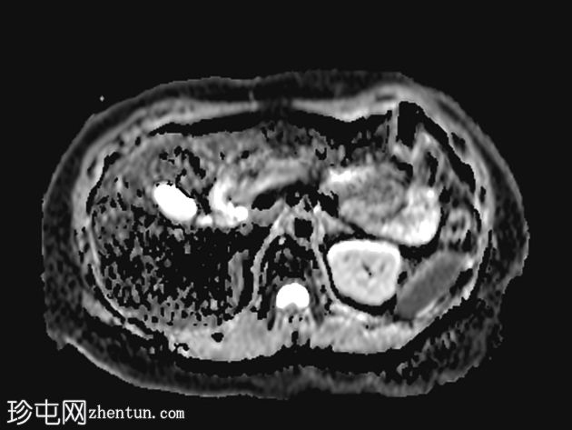
Axial
ADC
大壶腹部(内侧壁、下部 D2 段)区域增强中间 T2 信号团块,伴有轻度扩散受限。胰腺外观正常。胰腺分裂,主胰管通过圣托里尼管引流。无主胰管扩张。肝脏内有散在微小囊性病灶。
病例讨论
患者随后接受 EUS FNA 检查,最终诊断为胃肠道间质瘤 (GIST)。GIST 出血被认为是患者缺铁性贫血的原因。 |