介绍
非特异性上腹部疼痛。
患者数据
年龄:60岁
性别:女
CT
钙化肝包虫囊肿
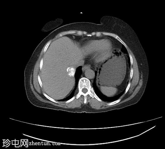
Axial
non-contrast
钙化肝包虫囊肿
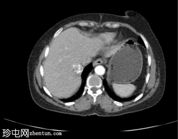
Axial C+
arterial phase
钙化肝包虫囊肿
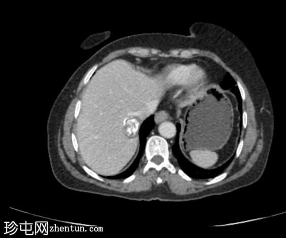
Axial C+ portal
venous phase
钙化肝包虫囊肿
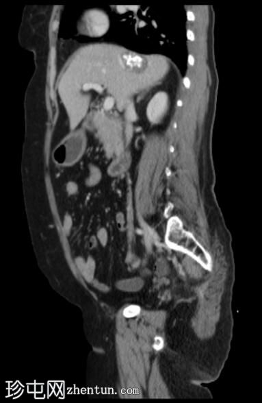
Sagittal C+ portal
venous phase
钙化肝包虫囊肿
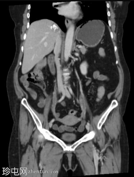
Coronal C+ portal
venous phase
钙化肝包虫囊肿
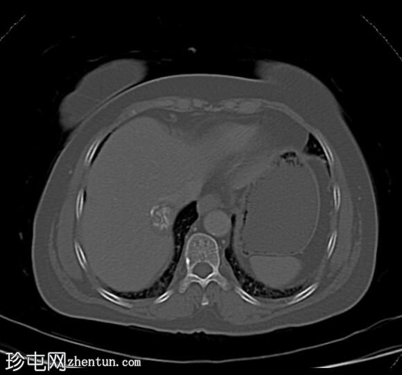
Axial bone
window
在右肝叶的包膜下区域观察到边界清楚的钙化病灶,具有多个内部匍匐状低密度线性结构,在所提供的相上没有任何密度变化,这很可能是钙化包虫囊肿。
肝脏位置正常,大小正常,边界光滑。
其余可视化的内脏看起来并无异常。
案例讨论
CT 结果与所描述的最有可能的钙化包虫囊肿有关。 |