介绍
无痛性血尿
患者数据
年龄:60岁
性别:男
CT
Bosniak IV 分类的肾囊性病变
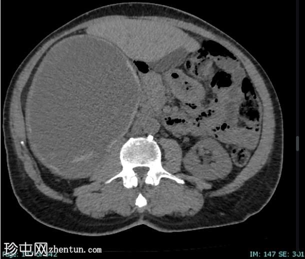
Axial
non-contrast
Bosniak IV 分类的肾囊性病变
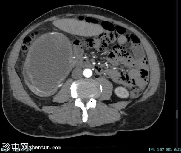
Axial C+
arterial phase
Bosniak IV 分类的肾囊性病变
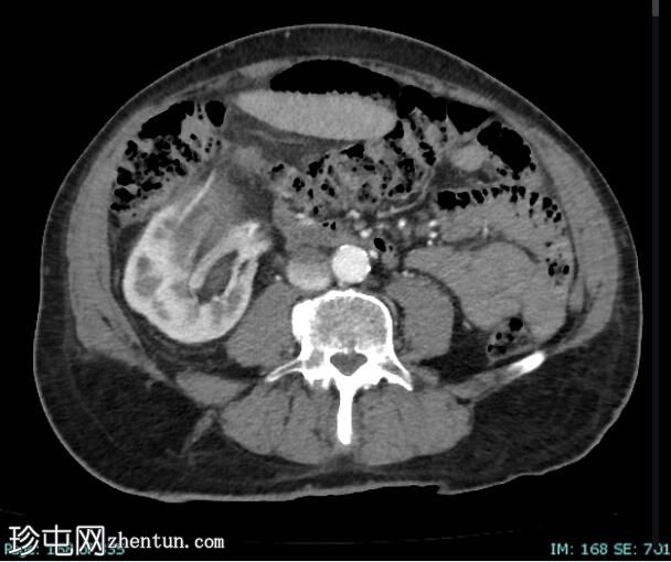
Axial C+ portal
venous phase
Bosniak IV 分类的肾囊性病变
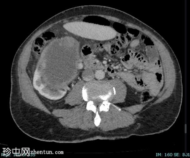
Axial C+ portal
venous phase
Bosniak IV 分类的肾囊性病变
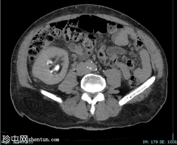
Axial C+
delayed
Bosniak IV 分类的肾囊性病变
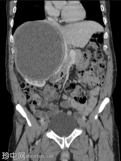
Coronal C+ portal
venous phase
右肾上部出现巨大的外生性囊性病变,对邻近的腹部结构产生占位效应,将其移位到左侧和下腔静脉(导致右侧精索静脉曲张)。 病灶壁较厚,内间隔较厚,钙化,边缘有少量强化结节。 囊肿下部有外部膨出,紧邻横结肠,无明显的脂肪劈裂线。 该囊性病变尺寸约为 15.2 x 19 x 18.5 厘米,周围有微妙的脂肪平面缠绕。
邻近的小椭圆形淋巴结尺寸为 1 x 2 厘米。
肾静脉开放。
右肾由两条动脉供血,对比之下显得不透明。
双侧肾少数有小囊肿。
案例讨论
Bosniak IV 囊性肿块具有超过 91% 的恶性病变风险。
治疗通常包括部分或全部肾切除术。
参考
1. Graumann O, Osther S, Osther P. Characterization of Complex Renal Cysts: A Critical Evaluation of the Bosniak Classification. Scand J Urol Nephrol. 2011;45(2):84-90. doi:10.3109/00365599.2010.533695 - Pubmed
2. Ferreira A, Reis R, Kajiwara P, Silva G, Elias J, Muglia V. MRI Evaluation of Complex Renal Cysts Using the Bosniak Classification: A Comparison to CT. Abdom Radiol (NY). 2016;41(10):2011-9. doi:10.1007/s00261-016-0797-5 - Pubmed
3. Graumann O, Osther S, Karstoft J, Hørlyck A, Osther P. Bosniak Classification System: A Prospective Comparison of CT, Contrast-Enhanced US, and MR for Categorizing Complex Renal Cystic Masses. Acta Radiol. 2016;57(11):1409-17. doi:10.1177/0284185115588124 - Pubmed
4. Bosniak M. The Current Radiological Approach to Renal Cysts. Radiology. 1986;158(1):1-10. doi:10.1148/radiology.158.1.3510019 - Pubmed
5. Skolarikos A, Laguna M, de la Rosette J. Conservative and Radiological Management of Simple Renal Cysts: A Comprehensive Review. BJU Int. 2012;110(2):170-8. doi:10.1111/j.1464-410X.2011.10847.x - Pubmed |