介绍
慢性咳嗽
患者数据
年龄:10 岁
性别:男
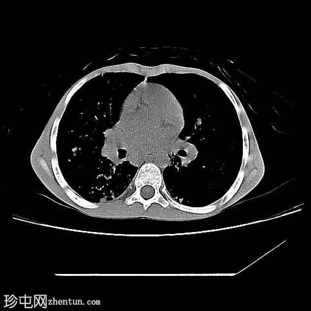
CT
囊性纤维化

Axial
non-contrast
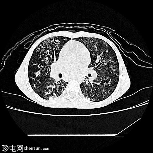
囊性纤维化

Axial lung
window
可见广泛的圆柱形和囊性支气管扩张,支气管壁增厚。
由于粘液堵塞,多个双侧小叶中心结节呈树芽状。
由于空气滞留,双肺马赛克衰减。
发现符合囊性纤维化的肺部表现。
病例讨论
囊性纤维化的肺部表现是最著名的表现之一。
HRCT 已成为 CF 患者监测中不可或缺的一部分,用于指导治疗和评估治疗反应,因为它不仅与肺功能测试相关,而且在某些情况下可以先发制人。通常每 6 至 18 个月重复一次扫描,具体取决于机构和临床病程。
参考文献
1. S. Karger AG, Karger. Cystic Fibrosis. (2001) ISBN: 9783805572248 - Google Books
2. David A. Warrell. Oxford Textbook of Medicine. (1985) ISBN: 9780198569787 - Google Books
3. Monroe E, Pierce D, Ingraham C, Johnson G, Shivaram G, Valji K. An Interventionalist’s Guide to Hemoptysis in Cystic Fibrosis. Radiographics. 2018;38(2):624-41. doi:10.1148/rg.2018170122 - Pubmed
4. Taylor-Cousar J, Robinson P, Shteinberg M, Downey D. CFTR Modulator Therapy: Transforming the Landscape of Clinical Care in Cystic Fibrosis. Lancet. 2023;402(10408):1171-84. doi:10.1016/s0140-6736(23)01609-4 |